CT Terminology and History
Objectives:
- Define terminology reviewed in this course
- List the different types of pitches
- Explain how collimation affects image quality and patient dose
- Understand the evolution of the CT Scanner
- Explain the principle of tomography
- Compare 2D, 3D, 4D, and 5D images
- Define Dual Energy CT scanning
Description:
The terminology utilized in CT is very different from the terms used in diagnostic radiography. Much of this can be attributed to the works of Dr. Godfrey Hounsfield and Dr. Allan Cormack who shared the Nobel Prize in medicine and physiology for their work in the invention and development of the CT scanner. Also, Computed tomography (CT) can be known as one of the most important diagnostic tools of the 21 st century. While this tool requires competent operators, each subsequent technological generation has required the technologist to be even more highly efficient. Therefore, this article will show the terminology used in CT, discuss the evolution of the CT scanner, and explain how these advancements have changed the process of CT. This article is accredited by the ASRT for 2 Category A CE Credits.
The terminology utilized in CT is very different from the terms used in diagnostic radiography. Much of this can be attributed to the works of Dr. Godfrey Hounsfield and Dr. Allan Cormack who shared the Nobel Prize in medicine and physiology for their work in the invention and development of the CT scanner.
Also, Computed tomography (CT) can be known as one of the most important diagnostic tools of the 21st century. While this tool requires competent operators, each subsequent technological generation has required the technologist to be even more highly efficient. Therefore, this article will show the terminology used in CT, discuss the evolution of the CT scanner, and explain how these advancements have changed the process of CT.
Targeting
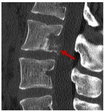
Targeting is a magnification technique done through reformatting. The reformatting process reconstructs image data in smaller pixel sizes to permit magnification with less distortion. This is done for areas where fine detail is needed, such as in the inner ear. Here you can see the magnified fracture in the lumbar spine.
Definition
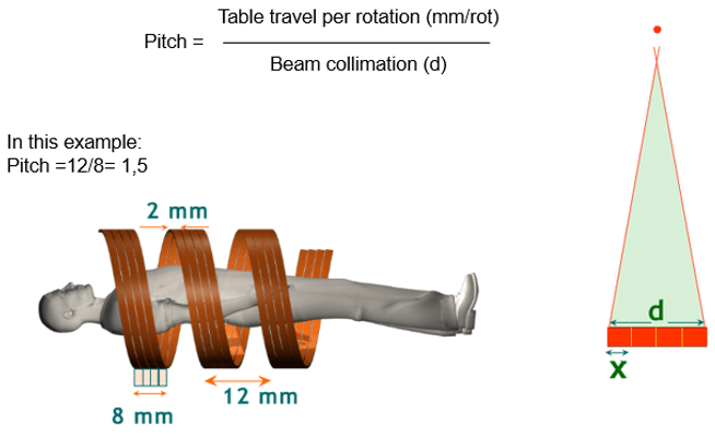
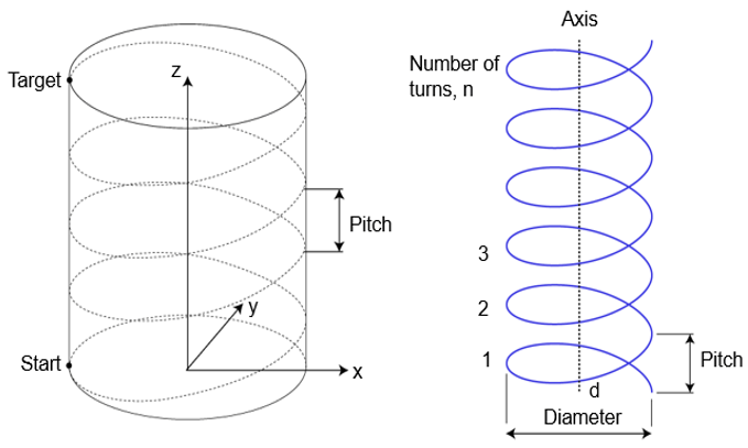
The pitch is defined as the table distance traveled for one 360 gantry rotation divided by the beam collimation. Pitch plays an important role in the image quality as well as patient dose.
Pitch Types
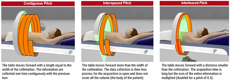
The 3 different types of pitch: contiguous, interleaved and interspaced.
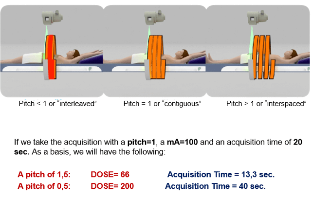
As shown in the image, higher pitch reduces image quality and patient dose while lower pitch increases image quality and increases the patient dose. For example, the image quality for a pitch of 0.5 is improved, but the patient dose is abnormally increased.
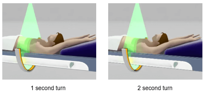
When we increase the time of the rotation speed of the detectors, the acquisition time is reduced but the image becomes noisier. This is due to the system’s inability to correct the maximum information per second.
Gantry
The gantry is a mounted framework that surrounds the patient. The gantry houses the x-ray tube, x-ray generator, detector systems, collimators, and the rotational frame all of which are considered the imaging components. Two important features of the gantry are the gantry tilting range and the gantry aperture. It can be tilted plus or minus 30 degrees. Most scanners have a 70cm aperture. The table flows into and out of the gantry during a CT examination.
Filtered Back-Projection
Also known as the filtered back projection, the projection profile is filtered or convolved to remove the typical blurring of the simple back-projection technique. Applying a filter function to an attenuation profile is called convolution. Filter functions may be referred to as algorithms, convolution filters, or kernels. Filter functions can only be applied to raw data.
Convolution
Convolution, a general-purpose algorithm, is performed on the reformatted data by the array processor. During convolution, the convolution kernel (a group of pixels) moves across the image pixel by pixel and is used to compute the value of the corresponding output pixel. Then the Back projection reconstruction algorithm reconstructs an image of the internal structures being scanned.
Back projection is the actual process of reconstruction to produce the image. The back-projection has two distinctive limitations, noise and streak artifacts. It is a combination of these restrictions and the advancement of computers that iterative algorithms have slowly replaced this method of image reconstruction.
Mask
It is sometimes useful to modify the value of each pixel to enhance the image. It is done through a mathematical filtering process. A set of mathematical operations is placed over each pixel, the pixel value becomes changed, and then the operation is slid over the next pixel. The total process is one of sliding the mask over all the pixel data and then displaying it as a modified image.
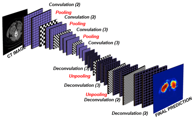
Deconvolution
Deconvolution is the process of returning the pixel values to their original level by a reverse process from convolution.
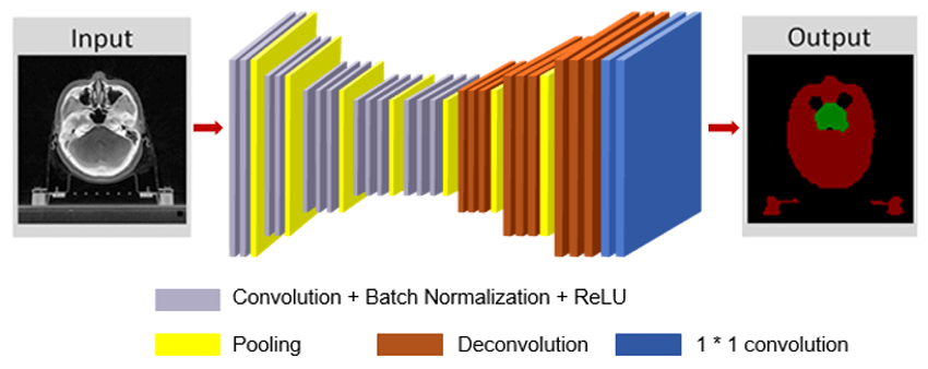
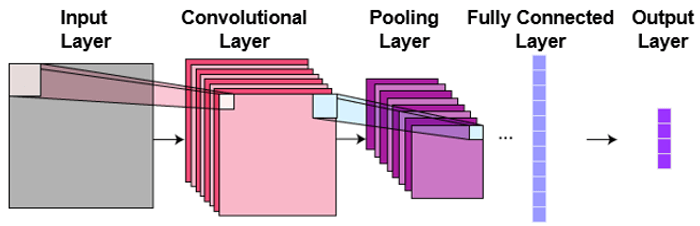
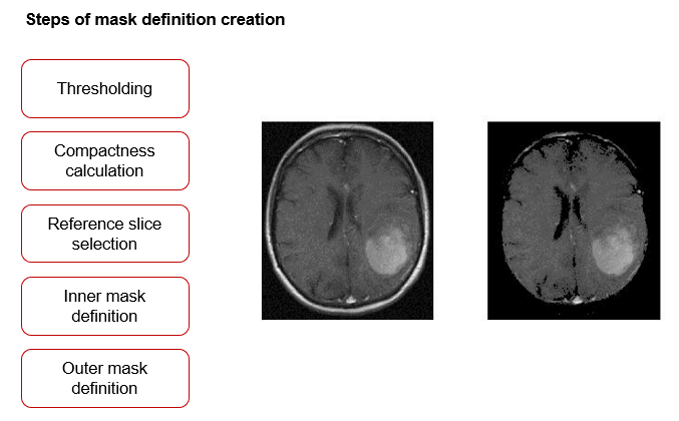
Automated Segmentation of Kidneys
The techniques available for the segmentation of medical images are specific to the application, imaging modality and type of body part to be studied. The automatic segmentation of medical images is a difficult task as medical images are complex and rarely have any simple linear features. Further, the output of the segmentation algorithm is affected due to:
- Partial volume effect
- Intensity inhomogeneity
- Presence of artifacts
- And closeness in gray level of different soft tissue
Helical Interpolation
Interpolation is a mathematical process used to smooth, enlarge or average images that are being displayed with more pixels than that for which they were originally reconstructed. Since helical scanning occurs in a pattern that is at an angle to the perpendicular desired, interpolation is used to straighten up the sections. This can be done through algorithms creating sections that are a reconstruction of data and not additional radiation to the patient.
Collimator
In CT, collimation is equally important as it affects image quality and patient dose. The basic collimator scheme in CT includes adjustable pre patient and post patient collimators along with pre detector collimators. In general, collimator sections are carefully arranged to shape the beam. Note that a Better recovery due to larger detector collimation.
Computed tomography can be known as one of the most important diagnostic tools of the 21st century. While this tool requires competent operators, each subsequent technological generation has required the technologist to be even more highly efficient.
- CT: Computerized Tomography
- CAT scan: Computerized Axial Tomography
- A CT scan makes use of computer-processed combinations of many X-ray images taken from different angles to produce cross-sectional (tomographic) images (virtual “slices”) of specific areas of a scanned object.
Volume
A volume is reconstructed from the axial images which are already acquired. Volume formation involves stacking images to form a volume with some preprocessing. Example : One page + one page + one page + … = One book. One axial image + one axial image + one axial image + … = Volume
Advantages
The principle advantages of CT are no superimposition of structures, better contrast resolution and third axial plane and 3D reconstruction.
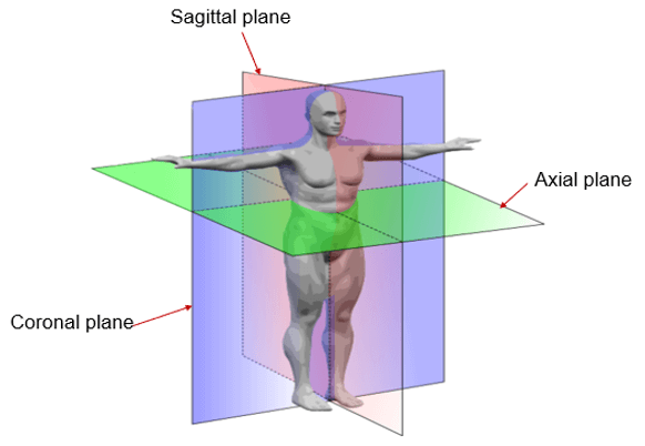
Anatomical Planes
Body planes are hypothetical geometrical planes used in medicine to divide the body into sections. They are commonly used in human anatomy to describe the location or direction of bodily structures. Reference planes are the standard planes used in anatomical terminology they include the sagittal plane, the coronal plane, and the transverse or axial plane. While these are the major reference planes of the body, other planes are commonly used in relation to these three.
General Components
The main components of a CT scanner are x-ray source and generator, collimator, detector array, data acquisition system, gantry and table, computational hardware and display monitors.
Table Limits
Some patients cannot be scanned because their weight limit exceeds that of the table’s capacity (generally in the range of between 400 and 500 pounds). The Toshiba Aquilion ONE has a capacity of about 660 pounds.
General Components Detectors
The detectors, which receive the transmitted x-ray beam after it has passed through the patient, convert the photons from the x-ray beam to an electrical signal, while the Data Acquisition System (DAS) converts the electrical signals from analog to digital. Detectors in a CT system should be small in size and be capable of responding with extreme speed to a signal.
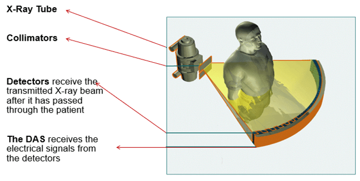
The Collimators
The tube collimator reduces patient dose and scatters radiation by restricting the volume of tissue that is being irradiated. This principle is the same as in diagnostic radiography. The tube collimation is pre-patient (restricting the tissue irradiated) and the detector collimator is post-patient collimation.
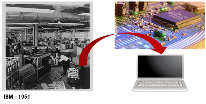
Technology for computed tomography very quickly advances after Godfrey Hounsfield’s work in 1970 – 1971, though in the early 60s preliminary work was being researched. Hounsfield along with Roentgens discovery of x-rays were two of the most outstanding contributions in medical imaging.
The original EMI head scanner was considered the first generation, but things progress rapidly after Hounsfield’s discoveries. Let us look at the generations of scanners over the years.
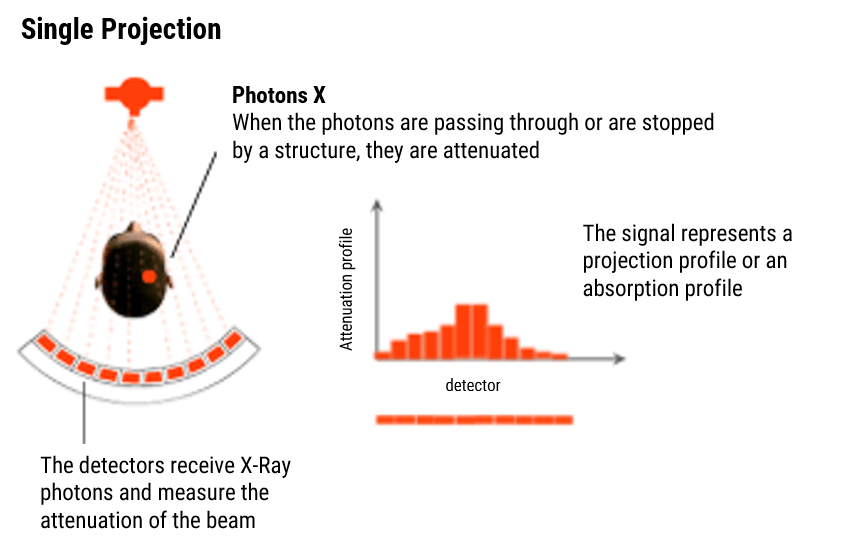
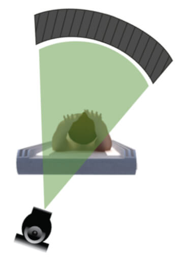
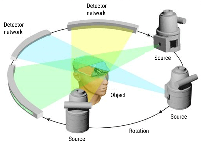
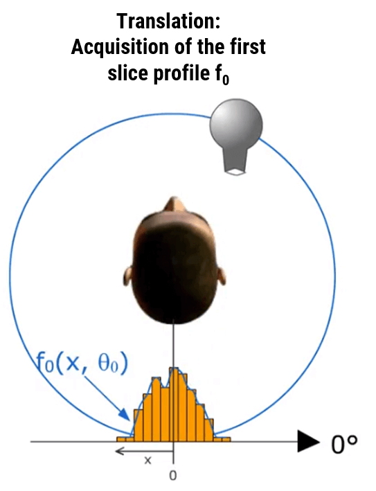
Principle of Tomography
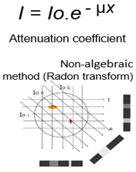
A CT scan or computed tomography scan makes use of computer-processed combinations of many X-ray measurements taken from different angles to produce cross-sectional images of specific areas of a scanned object, allowing the user to see inside the object without cutting. Let consider a parallel beam (for simplification). An appropriate linear attenuation coefficient is assigned to each pixel µi,j. Each detector (k) will measure the intensity Ik after propagation along each aligned pixels.
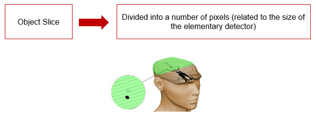
A CT image is typically called a slice, as it corresponds to what the object being scanned would look like if it were sliced open along a plane. So, while a typical digital image is composed of pixels (picture elements), a CT slice image is composed of voxels (volume elements).
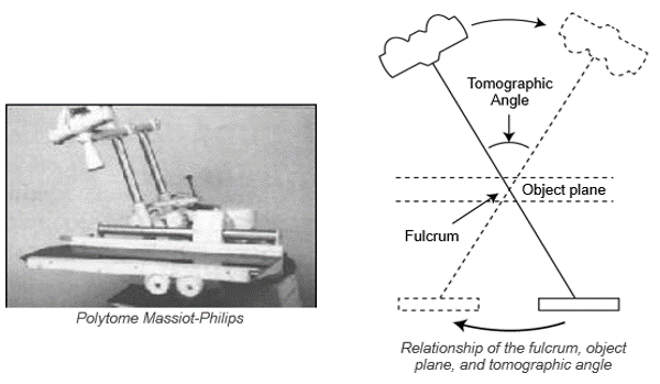
The tomographic movement is intended to isolate a plane within a volume by combining the movements of the tube and the image receptor. Tomography was used in diagnostic radiography often to visualize the layers of the kidney.
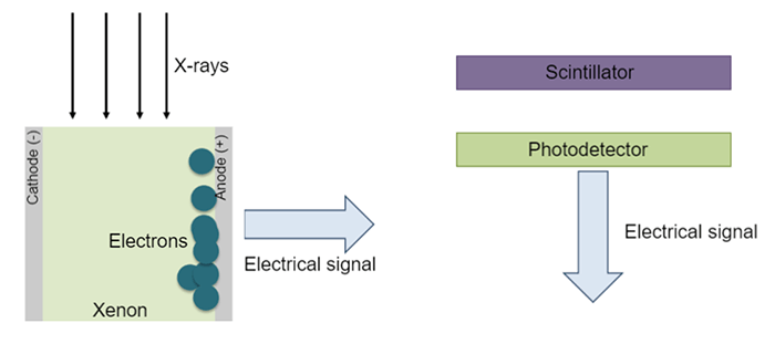
Detectors
Detectors should have a high capture efficiency, a high absorption efficiency, and a high conversion efficiency. Early scanners used a scintillation crystal which converted x-ray photons into light photons. Newer solid-state detectors use a scintillation material such as cadmium tungstate or a rare earth oxide ceramic bonded to a photodiode. Modern scanners use multiple rows of detectors.
When the x-ray beam interacts with the xenon gas, the gas is ionized. The electrons produced then travel and accumulate in the anode. This is them transmitted into an electrical signal which is sent to a computer system.
Xenon detectors
- High pressure xenon gas
- 10 to 20 bar
- 700 to 1000 cell measurements (flat electrodes)
- EDQ < 70%
Solid-state detectors
- Cadmium tungstate (example)
- Coupled to photodetector
- EDQ > 100%
First Generation
The first generation was the head unit only. It used a linear scan motion at each degree from 0-180* with rotation paired with a single detector. It took about 4.5 to 5 minutes of scan time per slice using a single ray pencil beam. Things advanced quickly from there.
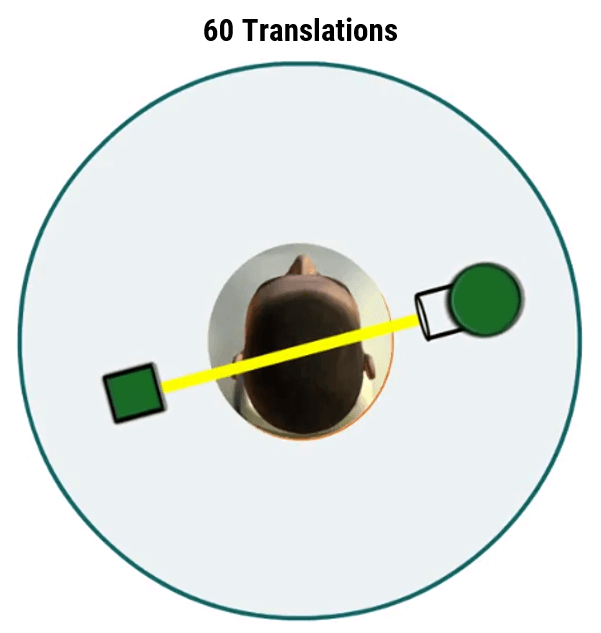
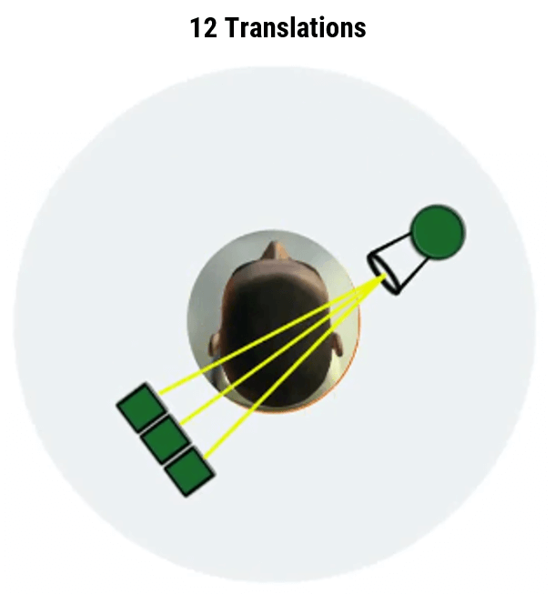
Acquisition Principle
In the first CT generation, the source-detector rotates slightly and a subsequent set of measurements are obtained during a scan (translate) and rotate (index) system.
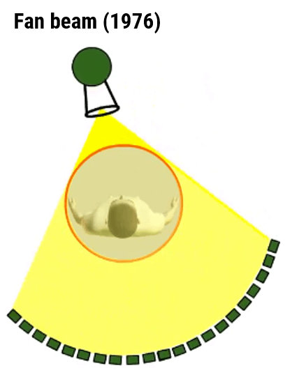
Second Generation
In the second generation, a single-projection, the narrow fan-shaped beam was used instead of a pencil slit. It had a linear array with as many as 30 detectors. Because of the loss of collimation, the amount of scatter radiation was increased. The scan time was reduced to about 20-90 seconds per slice which was short enough to allow imaging of the whole body.
Third Generation
In the third generation, huge modifications were applied. These units used a wider fan-shaped beam and a curved array of 250-750 detectors to acquire a single projection. The detectors could be collimated to reduce the amount of scattering. Scanning times were reduced to between 1 and 12 seconds allowing images to be produced sequentially to approximate dynamic functions, though in a crude manner.
Fourth Generation
As a result of a competitive contract awarded to the American Science and Engineering Company (AS & E), the fourth generation of CT scanners was developed. It used a wide fan beam and 600-2000 stationary detectors arrayed in a 360* ring and a continuous rotation. With the ring of detectors, the movement of detectors was eliminated. The scanning time for this generation was 1 second or less with some scanners capable of dynamic scans in the range of 15 scans per minute.

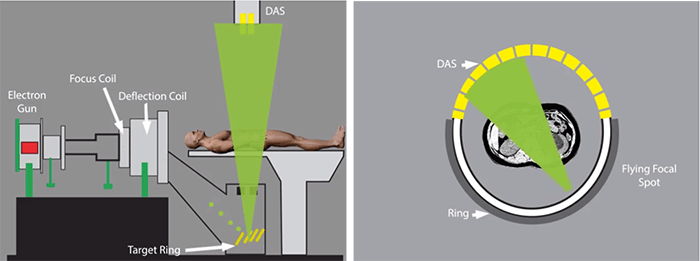
Fifth Generation
In the fifth generation, the x-ray tube is a large ring that circles the patient as opposed to the detector ring. This generation was used for cardiac tomography imaging and was referred to as Cine CT because the images were essentially anatomical movies. It was capable of 50msec scan time which could produce 17 slices per minute.
Sixth Generation
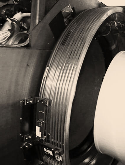
Helical scanners were made possible by advances in slip ring connection technology, Consisting of brushes that fit into groves, Slip rings permit the current and voltage to the tube to be supplied while the tube is in continuous rotation. Its primary advantage is a shorter scanning time which allows fewer contrast media to be used.
CT Slip ring technology was introduced to enable helical (continuous rotating) scanning. Before the introduction of Slip Rings, only Axial scanning was possible (which needed to stop/reverse the direction of rotation, after no more than 700 degrees rotation due to the finite length of the attached cables.) Slip ring technology eliminated the need for cables and enabled the continuous rotation of the gantry components.
Seventh Generation
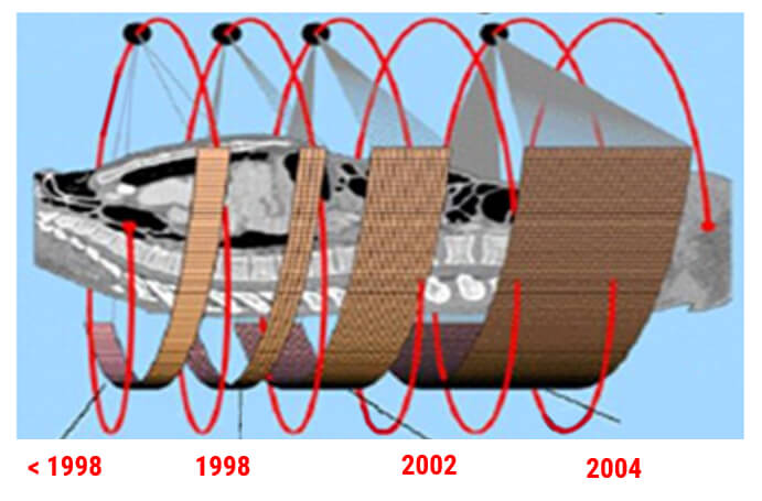
The seventh-generation introduced in 1998 gave us the most recent advancements. This uses usually 64-128 adjacent multiple detector arrays in conjunction with a helical CT scanner, the collimator spacing is wider and more of the x-rays that are produced by the tube are used in producing image data. Scanning time: 0.25 seconds.
Seventh generation ~ 1998
- Single X-Ray tube, Multiple-row detector, Rotating movement
- Allow simulataneous acquisition of multiple slice in a single rotation
- Half-second rotation (0.5s)
- Sub-second scanner
Dual Energy
By definition, dual-energy is the possibility of generating an image using two different energies. It can use a monotube with ultra-fast switching between low and high kVp ( 80 and 140) or bi-tubes with simultaneous acquisition using 2 tubes (80 kVp and 140 kVp or 100 kVp and 140 kVp).
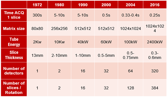
Evolution
Evolution of CT imaging over the years.
Evolution Table
When we look at this table, we see the evolution and comparison of CT advancements over the years as described in the explanations of the generations of CT scanners.
We can now begin to look at the differences in 2D, 3D, 4D, and 5D images as well as a combination of some of them and how they are used, We will discuss cinematic rendering and Hybrid 3D CT.
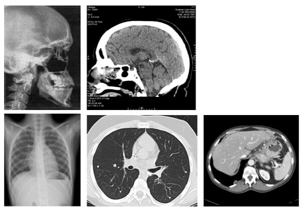
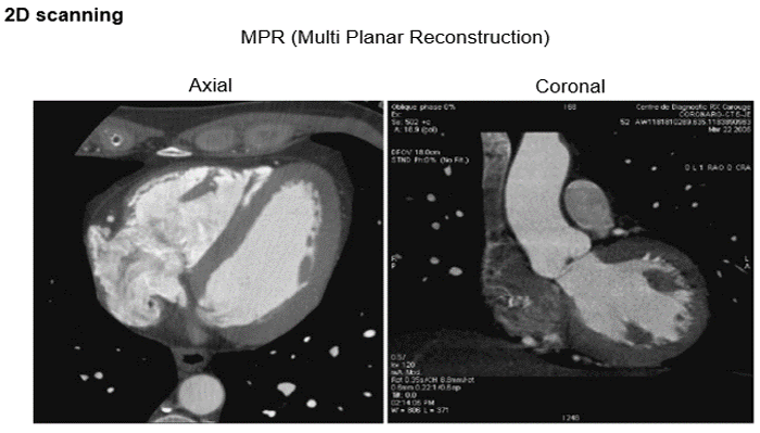
2 Dimensions
These images show us axial and coronal images in a 2D scanning mode.
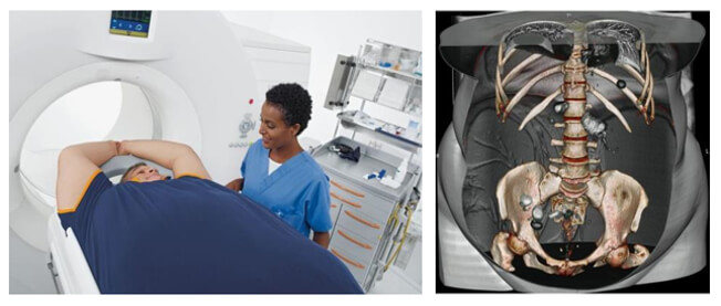
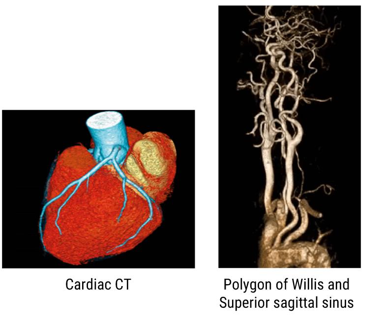
3 Dimensions
The image on the right side shows the Circle of Willis and the Superior sagittal sinus reconstructed using a volume rendering technique.
Hybrid 3D
These are a few examples of hybrid 3D imaging. All new scanners are now hybrid PET/CT scanners. The CT portion of these systems takes advantage of recent developments in multidetector CT technology.
In reality, PET/CT scanners are two separate machines in close proximity with a single table that moves the patient between the two. The PET and CT data are acquired sequentially.
4 Dimensions
4D scans show us movement in 3D images with time being the fourth dimension. The first model of the 256 slice 4D CT scanner was developed in 2003. In 2007, the 320 slice Aquilone One MSCT scanner was introduced at RSNA. This scanner can be used in neurological and cardiac imaging and other body applications where entire organs can be scanned very quickly.
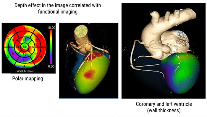
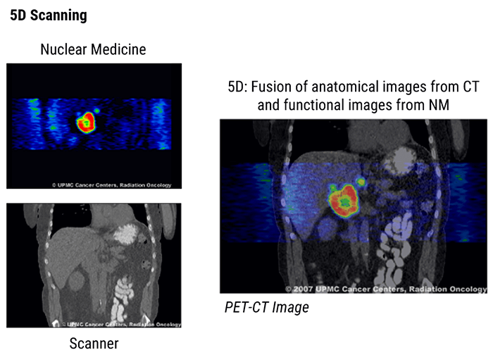
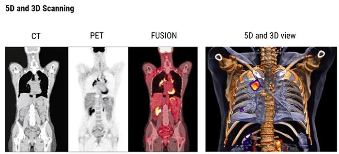
5 Dimensions
5D scanning (3D + dual-energy + time) is the fusion of anatomical images from CT and the functional images from Nuclear Medicine or MRI. Five-dimensional (5D) images are dynamic 3D images (4D) that are acquired at multiple times. A typical example would include dynamic cardiac CT scans and/or gated cardiac MRI acquired at 3-monthly intervals and dynamic cell growth and shape change in weekly intervals.
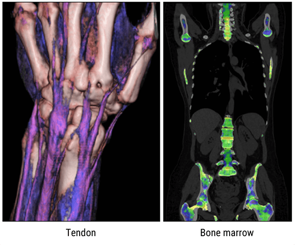
Examples
The below images show 3D scanning using the dual-energy technology.
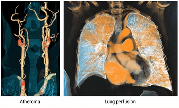
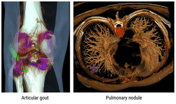
Additional examples highlighting articular gout as well as a pulmonary nodule using dual-energy technology.
Cinematic Rendering
Cinematic rendering is a novel post-processing technique for 3D visualization of CT image data. Compared to volume rendering, cinematic rendering results in a more photo-realistic representation of anatomy and provides the best image quality of high-density structures.
Post-Test & CE Certificate:
Add to CartPrice: $8.00
| ✔ | Approved by the ASRT (American Society of Radiologic Technologists) for 2 Category A CE Credits |
| ✔ | License duration: 6 months from purchase date |
| ✔ | Meets the CE requirements of the following states: California, Texas, Florida, Kentucky, Massachusetts, and New Mexico |
| ✔ | Meets the ARRT® CE reporting requirements |
As per the ARRT regulations, you have up to 3 attempts to pass the Post-Test with a minimum score of 75%.
Upon the successful completion of the Post-Test (score 75% or more), you will need to fill up a 1 min survey and then you will be able to issue your CE Certificate immediately.
Refund Policy: Non-Refundable
